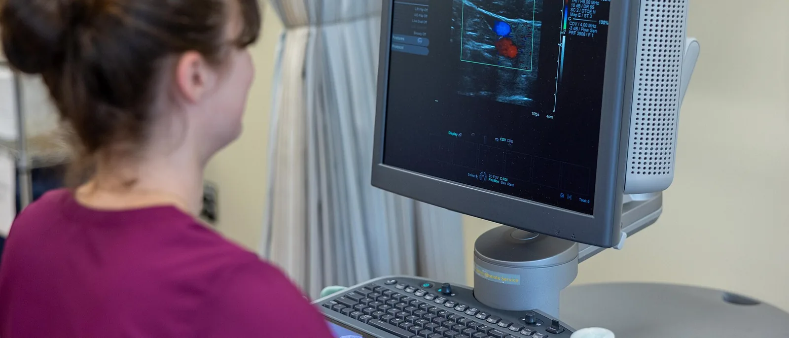
The standard heart ultrasound is a transthoracic echocardiogram, in which the probe is placed on the chest. You lie on your left side or on your back while the probe is placed on the left side of the chest and just below the breastbone, showing the heart as it beats along with the blood flowing through the valves. What some hospitals call a detailed heart ultrasound is usually the same transthoracic echocardiogram; a transesophageal echocardiogram, in which the probe is passed down the esophagus, or a stress echocardiogram, done while the heart is put under extra strain, is performed at a larger hospital.
The first thing to look at on a heart ultrasound report is the left ventricular ejection fraction, the share of blood the heart pumps out with each beat; a value of about 50% or higher generally means pumping function is preserved. A small amount of backward leakage through a valve, called trivial or mild regurgitation, is common in healthy people and usually needs no treatment. If the results show a thickened heart wall, reduced pumping function or a narrowed valve, we look into the cause and recommend a cardiology visit when needed.
For women, the heart ultrasound is done the same way. Because the probe is placed on the chest, you usually remove your top and bra and wear an exam gown that opens at the front, with only the area being scanned uncovered. It uses no radiation, so it can be done during pregnancy when needed.